


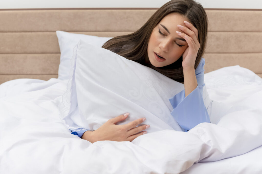
You may wake up unable to move while your mind feels fully awake. That sudden paralysis has a clear brain basis.
Sleep paralysis happens when the dream state of REM sleep overlaps with wakefulness, so your body stays “switched off” while your mind is alert.
We will walk through what the brain does during REM sleep and why that muscle paralysis sometimes lingers into waking. You will also learn which sleep habits and health issues make episodes more likely.
You will learn simple steps to lower the chance of an episode. We will also cover what to do if it happens.
Key Takeaways
- REM-related muscle paralysis can persist into wakefulness and cause sleep paralysis.
- Irregular sleep, certain health conditions, and sleeping on your back can raise your risk.
- Improving sleep routines and seeking care when episodes recur can reduce frequency.
What Is Sleep Paralysis?
Sleep paralysis is a brief state where the mind is awake, but the body cannot move. We will describe what it is, how it links to REM sleep and REM atonia, and the differences between isolated and recurrent forms.
Scientific Definition and Overview
Sleep paralysis is a type of parasomnia in which a person becomes conscious while voluntary muscles remain temporarily inactive. We experience awareness of the environment but cannot move or speak for seconds to a few minutes.
Episodes often include vivid perceptions or hallucinations, such as sensing a presence or feeling pressure on the chest. Researchers classify sleep paralysis as a REM-related phenomenon because it involves the same motor inhibition that occurs during dreaming.
It can affect people of any age and may happen once or recur. Risk rises with disrupted sleep, irregular schedules, stress, and certain medical conditions.
REM Sleep and REM Atonia
REM sleep is the sleep stage when most vivid dreaming happens, and brain activity resembles wakefulness. During REM, the brain triggers REM atonia—a neural shutdown of most skeletal muscles to prevent us from acting out dreams.
This atonia normally ends as we wake, restoring movement. Sleep paralysis occurs when awareness returns before REM atonia has fully lifted.
We are conscious, but the motor systems remain suppressed. That mismatch creates the core symptoms: immobility plus possible dreamlike hallucinations.
REM atonia is essential for safe dreaming, but its overlap with waking awareness produces the alarming sensation we call sleep paralysis.
Isolated vs. Recurrent Sleep Paralysis
Isolated sleep paralysis refers to single or occasional episodes that are not tied to another sleep disorder. We might have one episode in our life, often during a period of poor sleep or high stress.
Isolated cases usually need simple sleep hygiene changes to reduce risk. Recurrent sleep paralysis means episodes happen repeatedly over months or years.
We see higher recurrence with chronic sleep disruption, narcolepsy, psychiatric conditions, or inconsistent schedules. Recurrent cases may merit medical evaluation to check for underlying disorders and to consider behavioral or medical treatments.
The Neurological Mechanism of Sleep Paralysis
We focus on how REM motor inhibition stays active while awareness returns. We also look at which brain areas control that shift and which neurotransmitters push the system toward paralysis or wakefulness.
REM Intrusion and Motor Neuron Inhibition
REM sleep normally turns off motor neurons, so muscles stay limp. During sleep paralysis, that REM atonia “intrudes” into wakefulness.
We remain conscious, but motor neurons in the spinal cord stay suppressed, so we cannot move or speak. This atonia uses inhibitory signals carried by GABA and glycine onto motor neurons.
Those inhibitory signals block muscle activation even though the cortical circuits that produce awareness have reactivated. The mismatch—active awareness plus continued motor inhibition—is the core of REM intrusion.
Episodes often start at sleep–wake transitions, especially when sleep is irregular or brief. Microsleeps and abrupt shifts from REM to wake increase the chance that motor inhibition outlasts REM, producing the familiar frozen state.
Brain Regions Involved
The brainstem, especially the subcoeruleus (sublaterodorsal) region, drives REM atonia. It sends descending signals that activate inhibitory interneurons in the spinal cord.
If those brainstem signals persist while the forebrain wakes, paralysis continues. Forebrain areas like the thalamus and visual cortex restore sensory awareness.
The amygdala can amplify fear and produce strong emotional or hallucinatory content during episodes. Activity in these forebrain regions without coordinated brainstem shutdown creates the clash we experience.
Other modulatory centers, including hypothalamic orexin neurons, help stabilize wakefulness. When their input weakens, state control becomes unstable, and REM intrusion is more likely.
Role of Neurotransmitters
GABA and glycine produce the direct inhibition of motor neurons that causes atonia. We find high GABA/glycine activity during REM; if it lingers at wake onset, paralysis happens.
Acetylcholine promotes REM circuitry and can help trigger atonia. Elevated cholinergic tone at the wrong time favors REM intrusion.
Serotonin and norepinephrine normally rise with wakefulness and oppose REM; low levels of these can let REM features persist into wake. Dopamine modulates arousal and reward; disrupted dopamine signaling may affect sleep stability but is less central to atonia.
Overall, the balance among GABA, glycine, acetylcholine, serotonin, and other modulators determines whether motor inhibition ends with REM or continues into waking.
Triggers and Risk Factors
We see sleep paralysis most often when normal REM timing gets disrupted. Several clear factors raise the chance of episodes, and each affects REM sleep or how quickly we wake.
Irregular Sleep Schedule and Sleep Deprivation
Irregular bedtimes and not getting enough sleep are some of the top triggers. When we shorten sleep on some nights and binge on others, REM sleep fragments.
That increases the chance that awareness returns before muscle atonia ends. Shift work, late-night screen use, or pulling all-nighters push REM into odd parts of our sleep cycle.
Insomnia and frequent awakenings do the same by breaking REM continuity. Keeping a fixed bedtime and getting 7–9 hours most nights lowers risk because it stabilizes REM timing and reduces those fragmented wake-REM overlaps.
Stress, Trauma, and Mental Health
High stress and trauma make episodes more likely by raising arousal and changing sleep architecture. Anxiety and PTSD especially increase REM fragmentation and vivid dreaming.
That makes dream content spill into wakefulness more often. We notice people with chronic stress report more frequent and more intense hallucinations during episodes.
Treating the underlying mental health issue—therapy for PTSD or anxiety, and stress-reduction practices—helps reduce both episode frequency and the fear that often makes them recur.
Genetics and Family History
Family history matters. Twin studies and family reports suggest a heritable component to recurrent sleep paralysis.
Genes likely influence REM regulation and how easily our brain shifts between sleep and wake states. A family history doesn’t guarantee episodes, but it raises baseline susceptibility.
When genetic risk combines with poor sleep habits or stress, episodes become more common. Knowing a family pattern helps clinicians consider screening and targeted sleep-stabilizing strategies earlier.
Substance Use
Alcohol and some drugs alter REM sleep and raise the risk. Alcohol may suppress REM early in the night and then cause REM rebound later, creating REM fragmentation.
Certain antidepressants and stimulants can also disrupt REM timing. We advise avoiding heavy drinking close to bedtime and reviewing medications with a clinician if episodes increase.
Substance use that fragments sleep or changes REM intensity directly raises the chance of waking into REM atonia and experiencing sleep paralysis.
Associated Sleep Disorders and Conditions
We focus on conditions that most often appear with sleep paralysis and those that can be confused with it. This helps clinicians and patients spot warning signs, find the right tests, and choose effective treatments.
Narcolepsy and Related Disorders
We see sleep paralysis commonly in people with narcolepsy. Narcolepsy causes sudden sleep attacks and excessive daytime sleepiness.
It often causes cataplexy—brief muscle weakness triggered by strong emotions. These features help distinguish narcolepsy from isolated sleep paralysis.
Polysomnography and a multiple sleep latency test (MSLT) are the usual tests we use to confirm narcolepsy. Low hypocretin (orexin) levels in spinal fluid support the diagnosis in some cases.
Treating narcolepsy—using stimulant or wake-promoting medications, sodium oxybate, and strict sleep schedules—often reduces both daytime symptoms and frequency of sleep paralysis. Other REM-related disorders, like REM sleep behavior disorder, involve abnormal motor activity during REM and need different management.
We watch for overlapping signs so we can refer patients to a sleep specialist when necessary.
Night Terrors and Differential Diagnosis
Night terrors and sleep paralysis can both cause fear at night, but they differ in timing and awareness. Night terrors occur during non-REM deep sleep, often in the first third of the night.
People typically do not remember episodes and appear confused or frantic when awakened. In contrast, sleep paralysis happens at sleep onset or upon waking and comes with clear awareness and REM-like hallucinations.
We use the patient’s report, bed-partner observations, and sleep study data to differentiate them. Treatment for night terrors focuses on safety, sleep hygiene, and addressing triggers like fever or medications.
For patients reporting both types of episodes, we screen for stress, irregular sleep, substance use, and other sleep disorders to tailor a treatment plan.
Hallucinations and Sensory Experiences
We often wake into sleep paralysis with vivid sensory events that feel real. These events include visual, auditory, and bodily sensations rooted in REM physiology and threat responses.
Hypnagogic and Hypnopompic Hallucinations
Hypnagogic hallucinations appear as we fall asleep; hypnopompic hallucinations occur as we wake. Both happen during transitions when REM features—dream imagery and muscle atonia—overlap with wakefulness.
We see visual scenes, hear voices or humming, and feel tactile sensations like tingling or pressure on the chest. These experiences are usually brief, lasting seconds to a few minutes.
They stem from the brain’s dream systems remaining active while motor control is still suppressed. That mix explains why images can be dreamlike yet feel present and why we cannot move to check them.
Sleep loss, irregular schedules, and some sleep disorders raise the chance of these hallucinations.
Intruder and Incubus Sensations
Two common themes are intruder hallucinations and incubus (or “old hag”) sensations. Intruder hallucinations present as a sensed presence, footsteps, whispers, or visible figures in the room.
Incubus sensations involve pressure on the chest, choking, or the feeling of being pinned down, often paired with intense fear. Both likely come from a mix of REM-related paralysis, altered breathing patterns, and activation of threat circuits in the midbrain.
The brain interprets restricted breathing and helplessness as an external attack, producing vivid threat imagery. We should note that while terrifying, these sensations are not signs of physical harm and usually stop when normal sleep–wake balance returns.
Cultural Interpretations
Cultures name and frame these experiences differently: “sleep paralysis demon,” “old hag,” “jinn,” or “incubus.” Those labels shape how people describe episodes and how much fear they feel.
Cultural stories can intensify the content of hallucinations by providing ready-made images—an evil figure, a suffocating presence, or a visitor. At the same time, knowing the sleep-based cause can reduce distress.
We can use cultural context to better understand a person’s reaction and to guide reassurance and practical steps, like improving sleep habits and seeking medical evaluation if episodes are frequent.
Is Sleep Paralysis Dangerous?
Sleep paralysis does not stop breathing or cause physical harm, but it can leave people shaken and affect sleep quality. We explain the likely physical and mental effects and when to get evaluated by a sleep specialist.
Physical and Psychological Impact
Physically, sleep paralysis affects voluntary muscles only; breathing and heart function remain normal. Episodes last seconds to a few minutes and end on their own.
People may feel chest pressure, shortness of breath, or tingling, but medical measures like oxygen or CPR are not needed. Psychologically, episodes can trigger intense fear, anxiety, and trouble sleeping afterward.
Recurrent events can cause anticipatory anxiety—worry about falling asleep—which raises insomnia risk. Vivid hallucinations during episodes can feed trauma-like distress, especially if culturally framed as supernatural.
Tracking episodes, sleep times, stress, and medications helps identify triggers and reduce frequency.
When to See a Sleep Specialist
We recommend seeing a sleep specialist if episodes are frequent, severely distressing, or paired with daytime sleepiness. A specialist will review sleep history, mental health, medications, and possible triggers such as shift work or irregular sleep.
Polysomnography (sleep study) may be ordered when symptoms suggest narcolepsy, sleep apnea, or other sleep disorders that fragment REM sleep. The study records brain waves, breathing, and muscle tone to pinpoint REM dissociation.
Treatment focuses on stabilizing sleep schedules and treating underlying disorders. When needed, brief medication or therapy may be used to reduce anxiety and improve sleep.
Managing and Preventing Sleep Paralysis
We focus on clear actions you can take to reduce episodes: improve nightly habits, use evidence-based therapy and relaxation skills, and consider medical options when needed. Each path targets specific triggers and can be combined.
Sleep Hygiene and Regular Sleep Schedule
We set a consistent bedtime and wake time, even on weekends, to stabilize REM cycles. Aim for 7–9 hours nightly and keep lights dim at least 30 minutes before bed to cue melatonin.
Avoid caffeine after mid-afternoon and heavy meals within two hours of sleep. Limit alcohol; it fragments REM and can raise risk.
We arrange the sleep space: cool temperature, low noise, and a comfortable mattress. Remove screens or use blue‑light filters an hour before bed.
If we nap, we keep naps under 30 minutes and before 3 PM to avoid shifting nighttime sleep. We track patterns for 2–4 weeks.
Noting when episodes occur helps link them to late nights, shift work, or irregular sleep.
Cognitive Behavioral Therapy and Relaxation Techniques
We use cognitive behavioral therapy for insomnia (CBT-I) to fix beliefs and habits that disturb sleep. CBT-I gives structured steps: stimulus control, sleep restriction, and cognitive restructuring.
These steps reduce wakefulness and REM instability. We add progressive muscle relaxation and breathing exercises before bed to lower arousal.
Tense-and-release of muscle groups for 10–15 minutes reduces the chance of waking into REM at a high-stress state. Mindfulness meditation for 10 minutes nightly helps reduce anxiety that often precedes episodes.
When anxiety or trauma play a role, targeted CBT or trauma-focused therapy can reduce recurrence by lowering nighttime hypervigilance.
Medical Treatments and Medications
We consult a sleep specialist when episodes are frequent, cause severe anxiety, or coincide with daytime sleepiness.
The clinician may order a sleep study to check for narcolepsy or sleep apnea.
If an underlying disorder shows up, treating it often cuts episodes.
For some patients, low-dose antidepressants that suppress REM—such as certain SSRIs or SNRIs—reduce sleep paralysis frequency.
We consider risks and side effects before use.
Short-term sleep medicine can help reset sleep patterns.
We discuss medication choices, dosage, and monitoring with our provider.
Understanding the Cause and Reducing Future Episodes
Sleep paralysis can feel alarming because your mind is awake while your body remains temporarily “switched off.” The science behind it is rooted in REM sleep: during REM, the brain naturally limits muscle movement (REM atonia) to prevent acting out dreams. Sleep paralysis occurs when that REM-related muscle inhibition overlaps with wakefulness—so awareness returns before the body’s normal movement fully comes back. This mismatch can also explain why some people experience vivid sensations or hallucinations during an episode, especially when sleep is fragmented or stress is high.
For many people, reducing episodes comes down to stabilizing sleep and lowering triggers that disrupt REM timing. Irregular sleep schedules, sleep deprivation, sleeping on your back, stress, and certain health conditions can all raise risk. Consistent sleep routines, improved sleep hygiene, and relaxation techniques can make episodes less frequent. However, recurrent sleep paralysis—especially with excessive daytime sleepiness or symptoms suggestive of narcolepsy or sleep-disordered breathing—warrants professional evaluation. Identifying and treating underlying conditions often provides the most reliable long-term relief.
At Gwinnett Sleep, our board-certified sleep specialists use advanced diagnostics and personalized treatment plans to evaluate recurring sleep paralysis and related sleep disorders. If episodes are frequent, distressing, or affecting your ability to sleep comfortably, our team can help you find answers and reduce future risk.
Schedule your consultation today and start sleeping the difference.